Ozone therapy is a versatile term that describes a practice in which ozone is administered to human body system via gas, water or oil. Potential benefits obtained are based on the principle that ozone inactivates disease microorganisms, improve cellular function, and promote the healing of damaged tissues.We present a case of ozone therapy used in form of ozonated oil on exophytic fibrous gingival lesion.
Methods
A 42-year female patient was selected presenting with mild to moderate painful, exophytic, fibrous lesion on upper anterior gingiva. Gingival lesion was treated with 2ml of ozonated oil, thrice daily for one week. After ozone therapy, postoperative outcomes were measured and analysed. Finally, the lesion was subjected to excisional biopsy and histopathological evaluation.
Results
After ozone therapy patients revealed less pain. On examination of lesion, there was improvement in clinical sign of inflammation, reduction in surface ulceration. During final biopsy, less bleeding was observed. Planimetrical analysis showed reduction in size of lesion. The histopathological analysis showed reduction in collagen fibers and inflammatory cell in connective tissue stroma.
Conclusion
Topicalozone therapy provided potential benefits to treated exophytic gingival lesion. Observed benefits in present case report needs to be verified in future well-controlled clinical trials.
INTRODUCTION
Ozone (O3), an allotropic form of oxygen is becoming increasingly important both in environmental context and in medical science. In the medical science, ozone has been shown to possess unique properties, which are being defined and applied to biological systems as well as to clinical practice of dentistry and medicine 1,2
Ozone therapy can be defined as a versatile bio-oxidative therapy in which oxygen/ozone is administered via gas or dissolved in water or oil base to obtain therapeutic benefits in a diseased living system.
Now a day’sozone gas dissolved in oil base is gaining importance due to its excellent curative results, simple application, longer-term effect and non toxic in nature3,4. In the past years, therapeutic effect of ozonated oil was attributed to its antibacterial, antifungal, antiviral, antiparasitic, antihypoxic, analgesic and immunomodulatory effects on biological systems 1,5
Recently topical ozonated oil application on human and animal models have been shown to improve cellular function, improve healing of tissue, and scavenge the defective tissue 3,6 in the biological system, promoting the healthy cells to survive and multiply more rapidly. Rationale behind these applications has been linked to action of ozone on biological activities occurring in a living system 7,8.
†Department of Periodontology,
JSSDentalCollege& Hospital Mysore-15 Karnataka, India
††Department of Oral Pathology and Microbiology,
JSSDentalCollege& Hospital Mysore-15 Karnataka, India
In the present context of time, these empirical mechanisms of actions are supported with few published case reports and scientific studies, which restrict the application of ozonated oil in different fields of medicine and dentistry.
With the few published and anecdotal information on ozonated oil and its therapeutic effect on biological tissues. We designed a therapeutic approach to an exophyticfibrous enlarged lesion on human gingiva. As a consequence, a human case was selected presenting with exophytic gingival lesion (indicated for excisional biopsy) towhich ozonated oil can be applied and further Planimetrical and histopathologicalevaluation can be made. Thus, an experimental attempt was made to provide initial information about the Planimetrical and histological changes occurring after topical ozonated oil application on an exophytic gingival lesion.
CASE REPORT ANDMETHODOLOGY
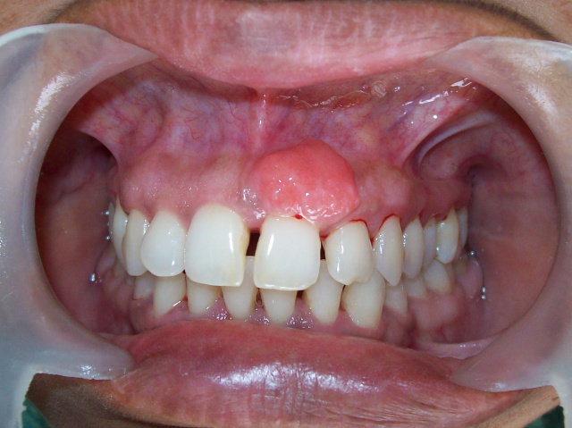
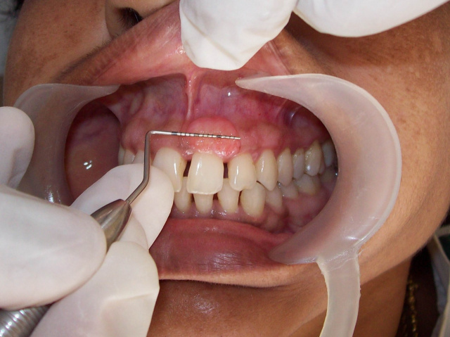
We selected a 42-year-old female patient who was referred to our department (Department of Periodontology, JSSUniversity, Mysore, India) with a chief complaint of a enlarged lesion on her upper anterior gingiva (Fig.1). Patient revealed that initially the lesion was small, but gradually increased in size since past 6 months. The lesion was mild to moderate painful during brushing or mastication. The lesion was not associated with pregnancy or any other systemic outcomes. Patient gave history that she was systemically healthy and had neither taken any medications like antibiotics and analgesics in the past 6 months, nor taking any systemic drugs, which has manifestation on the gingival tissue. On Intraoral examination, an enlarged lesion was seen, confined to attached gingiva of maxillary left central and lateral incisors. The lesion was 10 × 14 mm in size, exophytic, pedunculated, roughly oval, reddish, and ulcerated. On palpation, the lesion was firm and fibrotic in consistency, with bleeding on provocation. The overall oral hygiene status was good with minimal deposition beneath the lesion or around the associated teeth.
A treatment plan was designed which included, therapeutic application of ozonated oil followed by excisional biopsy and histopathologic evaluation as a diagnostic and therapeutic approach to treat the lesion.
The use of topical ozonated oil on oral diseased tissue was approved by the ethical committee of JSSUniversity, Mysore, India. A written informed consent was obtained from the patient for publication of this case report with the accompanying images. All procedures mentioned in this paper were performed according to the ethical principles established by the Declaration of Helsinki 9
Patient was scheduled on recall appointments. As a part of the routine procedure, a complete blood examination was made and further procedures were carried out only after confirmation of all normal haematological values. Furthermore, an intraoral periapical radiograph (IOPAR) of associated tooth/ lesion was taken to rule out any bony or periapical pathology associated with the tooth structure.
On the initial appointment no scaling or mechanical debridement was done. Only relevant medical history and complete dental history was recorded accompanied by a thorough clinical examination of the subjected lesion. Preoperative photographs were taken for image analysis.
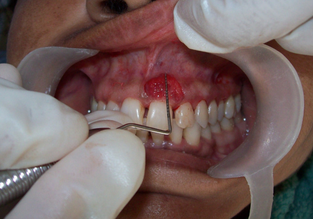
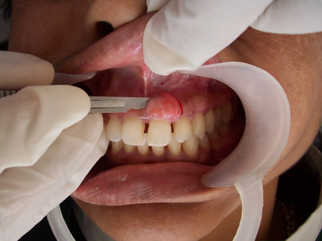
Next one-week appointment was scheduled for implementing the therapeutic application of ozonated oil and training the patient how to apply the oil on enlarged lesion. Before implementing therapeutic application of ozonated oil on the lesion, a small section of tissue mass measuring 9.8 ×3.2 mm was excised from the distal margin of exophytic gingival lesion under local anesthesia (2% lignocaine, 1:80000 adrenaline) (Fig.2). The obtained sectioned specimen was submitted to histopathological analysis. This was done in an attempt to take a control from the same lesion with which final histopathological outcome obtained after application of ozonated oil, can be compared.
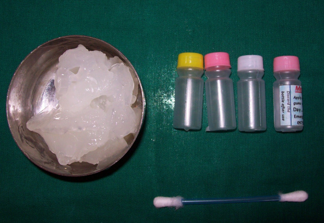
Two ml of ozonated olive oil estimated at 80µg/ml was applied on the lesion. For home use 21 premeasured disposable plastic vials, containing 2 ml of ozonated olive oil was given to patient(Fig.3) Patient was advised to apply 2 ml of ozonated oil with sterile cotton bud on lesion site three times daily for 7 days after meals. No antibiotics and/or analgesics were prescribed along with prescribed ozonated oil.
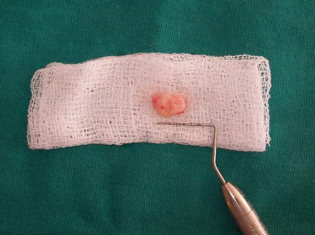
On the next, one-week recall visit patient’s centred clinical outcome, like pain and discomfort were recorded. Photographs of the lesion were taken for image analysis. Subsequently patient was prepared for final biopsy procedure. A measured volume of ozonated oil (0.5 ml) was applied on the lesion followed by complete excision of gingival lesion under local anesthesia (2% lignocaine 1:80000 adrenaline).The excised tissue specimen (Fig.4) was formalin fixed andstained with haematoxylin and eosin stain for the histopathological analysis. After final biopsy procedure the patient was again re-scheduled for next week appointment and further evaluation of post-excised wound on gingival surface was made.
For the each images taken at each time intervals computerised Planimetrical analysis was done to evaluate the difference in morphological size of lesion at each visit. At the each visit image of the lesion with overlying periodontal probe (UNC-15 Probe) was captured using a digital camera (Kodak C713, Eastman Kodak Company, Rochester, NY14650). Camera was kept at a distance of ≈ 25 cm and perpendicular to the subjected lesion. A set of 50 images were captured and reproduced images were analysed by image analysis software (UTH SCSA Image Tool; IT Version 2.0). Each image was analysed 20 times and the generated mean area (mm2) of measurement (Mean± SD) was considered for detecting the change in morphology of the lesion. In order to eliminate the discrepancy in measurement, that might have occurred due to lesion and camera distance. First, the scale was set in image analysis software by measuring a known distance of overlying periodontal probe markings. Subsequently the underlying lesion was measured using the same scale setting. This procedure was repeated for each reproduced image. Each lesion was measured after setting a scale from the overlying periodontal probe. Thus, a Planimetrical method with standardized scale setting technique was generated that eliminated the error that might have occurred during measurement of lesion. The obtained Planimetrical data of pre-ozone treated lesion and post-ozone treated lesion were compared and the difference was considered as change in morphological size of the lesion.
Results
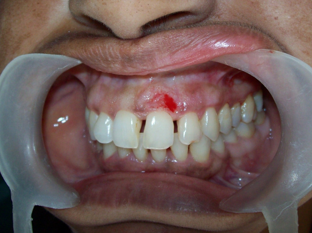
After one week, application of ozonated oil on gingival lesion clinical picture demonstrated a well-localized, slightly shrunk gingival mass, which was less firms on palpation. Surface foci of ulcerations were greatly reduced with less sign of acute inflammation on the surface when compared to previous pre-ozone treated lesion (Fig.5).
During the final biopsy procedure of post-ozone treated lesion, a dramatic reduction in bleeding was observed when it was compared to previous experience of bleeding during excision of marginal gingival specimen taken for control (Fig.6, 7).
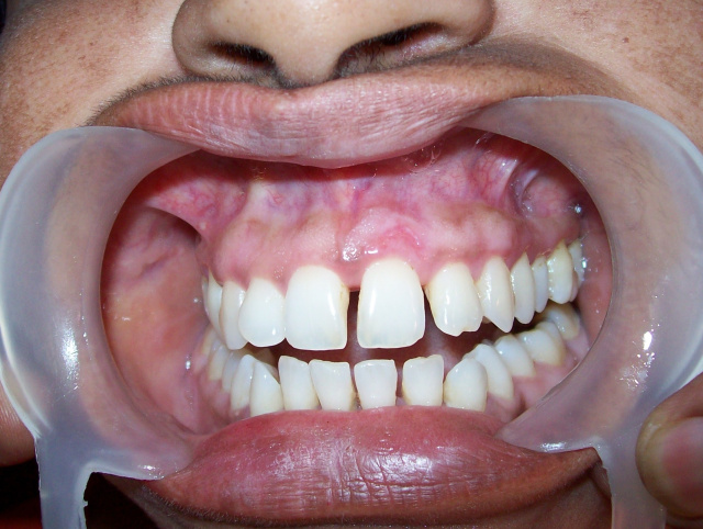
After complete excision of tissue, the final post-excised wound, healed uneventfully and revealed no sign of scarring or other adverse outcomes (Fig.8).
Planimertical changes revealed a decrease in mean area of gingival lesion. Original lesion was measured to be 140 mm2. After excision of control specimen (31.36 mm2), the remaining size of lesion was 108.64 mm². After one week of ozone application, the lesion size was 96.35mm² that demonstrated a reduction of 12.29 mm2 in ozonetreated lesion.
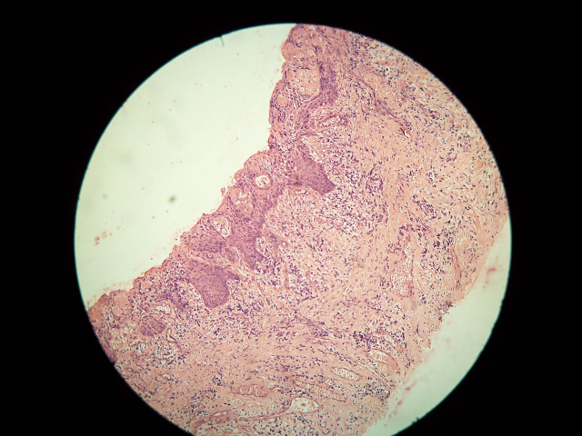
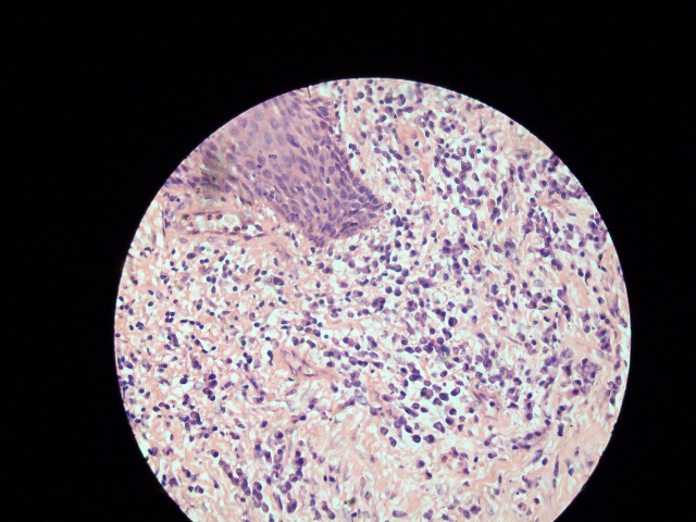
Histopathological examination of pre-ozone treated specimen revealed stratified squamous epithelium with multiple foci of surface ulceration. Connective tissue stroma showed thick collagen fibers bundles with plump fibroblast cells. Dense chronic inflammatory cells were evident, consisting of plasma cells, lymphocytes, and mast cells. Few blood vessels were also seen in connective tissue stroma. From the observed histopathological features, a diagnosis of inflammatory fibrous hyperplasia was confirmed (Fig.9a, 9b).
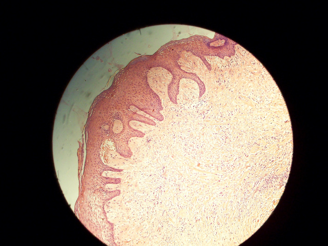
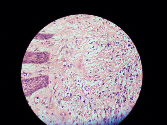
Post-ozone treated specimen revealed well-keratinized stratified squamous epithelium. There were fewer foci of surface ulceration with regenerating new epithelial cells covering the ulcerated areas. Connective tissue stroma showed less collagen fibers bundles with fewer fibroblast cells. There was a remarkable reduction in number of chronic inflammatory cells in the connective tissue stroma (Fig.10a, 10b).
DISCUSSION
This paper reports a case of ozone therapy used in form of topical ozonated oil on exophytic enlarged gingival lesion and its therapeutic benefits obtained after short-term applications.
Ozone therapy is a recognized, versatile, modern, bio-oxidative therapy known today to human civilization. The rationale behind bio-oxidative therapies is that, as long as the body's needs for antioxidants are met, the use of certain oxidative substances will stimulate the movement of oxygen atoms from the bloodstream to the cells 10. Moreover, ozone brings about the rise of pO2 in tissues and improves transportation of oxygen in blood, which results in change of cellular metabolism, activation of aerobic processes (glycolysis, Krebs cycle, β-oxidation of fatty acids) and use of energetic resources 11. It also prevents the formation of erythrocytes aggregates and increases their contact surface for oxygen transportation12. Thus, with higher levels of oxygen in the tissues, microorganisms are killed along with defective tissue cells resulting to healthy cells survive and multiply more rapidly.
In the present paper, patient centred clinical benefits were observed after one-week application of ozonated oil on enlarged gingival lesion. Patient reported with less pain and discomfort, improved clinical inflammatory sign, and reduced ulceration on the overlying epithelium of lesion with improved sign of healing. The exact mechanism behind it is still unclear.
Few authors 3,4,13 have proposed that base oils (Olive oil, Sunflower oil, Coconut oil, etc.) during ozonation process traps O3 in the form of a stable ozonide. Thus the produced stable ozonide when encounters the warm exudates of the wound, slowly decomposes to reactive ozone. Which readily dissolves in water, generates hydrogen peroxide and lipoperoxides that can explain the prolonged disinfectant and stimulatory activity on ulcerated chronic wounds.4 Moreover, the phenomenon of improved wound healing after ozone therapy has been related to the rapidly changing cell types and to the release of cytokines that modulate the complex healing process 14-18
Reduced inflammatory sign can be attributed to oxidizing potential of ozone 19. Ozone has been shown to oxidize prostaglandins20. Prostaglandin is a known biologically active compound that participates in inflammatory reactions, thus the local application of ozonides may promote removal of inflammatory phenomena.
Reduced pain can be related to membrane-stabilizing effect of ozone on pain receptor cells. Ozone has potential to increases the excitability threshold of pain receptor cell membranes 21 Direct oxidation of proteins (algopeptides) is another mechanism, which may be responsible for reduction in pain 22-24. Algopeptides are protein formed in the place of tissue damage and helps in transport of pain impulses to the brain.
There are some evidences, which reports, local application of ozonised oil significantly reduces pain in patients presenting with alveloitis when compared with application of control medications 25,26.Furthermore an evidence of ozonated oil in the treatment of oral apthous ulcer and alveloitis has been documented27. In this study, two groups of patients(n=142) presenting with oral aphthae and alveolitis were advised to apply 2 to 3 drops of the ozonized oil three times a day for five day, to their respective lesions. The result outcome revealed significantly reduced pain and better postoperative results in ozonated oil group compared to control groups. Our paper result is similar to these results where reduced pain has been reported after application of topical ozonated oil.
There is evidence 16 where, accelerated human oral wound healing with significantly improved postoperative outcomes after application of ozonated water has been documented. Application of ozonated water, accelerated the wound healing in the oral cavity within the first 48 hours compared to placebo water, resulting in earlier epithelial wound closure after 7 days. Our paper supports the findings of A.Filippi where improved healing was found after one week of ozone application.
The antimicrobial efficacy of ozonated oil is another mechanism, which can be attributed to improved patient centred clinical outcomes. Various in vitro and in vivo studies28-32 have revealed that ozonized oil when encounters a microorganism, a severe alteration in the cytoplasm of microbial cells occurs consequently leading to membrane rupture and leakage of cytoplasmic content.
During final excision of post-ozone treated enlarged lesion, a dramatic reduction in bleeding was observed.However, the exact mechanism is not clear, but could be ascribed to haemostatic changes that have been explained in a previous study.33 This in vivo study indicated that the arrest of bleeding under the influence of ozone is due to the formation of a fibrin membrane on the surface of the flowing blood, leading to rapid and effective haemostasis. These haemostatic changes have also been attributed to increased releases of growth factors after ozone treatment, consequently leading to vasoconstriction and platelet aggregation 34,35.
Previous study done by A Fillipi 36 have concluded that the ozonized water could act as a therapeutic agent in oral surgery raising haemostasis, enhancing oxygen supply and inhibiting bacteria proliferation. The author suggested that ozonized water could be employed during oral surgical procedures. Similar report is also from a study done during cardio-thoracic surgery where intraoperative use of ozone during surgical treatment effectively stimulated the system of haemostasis 37
Planimetrical analysis revealed a reduction in mean area of gingival lesion after application of ozonated oil. A reduction in size from 108.64 mm² to 96.35mm² with mean difference of 12.29 mm2 was observed. The exact mechanism behind the shrinkage of enlarged lesion is still unclear. The reason could be ascribed to reduction in size is the reduction in collagen fibres bundles and inflammatory cells infiltrate in connective tissue stroma. These histopathological changes were confirmed by histopathological examination of the sectioned lesion after ozonated oil application. Enhanced production of cytokines might have stimulated the fibroblast cell to activate phagocitary action on excessive collagen fibers lying in the connective tissue stroma. Another reason could be ascribed to the activation of local antioxidant defences mechanism by local ozone application, which might have stimulated wound contraction and approximation of wound edges created due to sectioning of control specimen from enlarged lesion.
Histopathological analysis of post-ozone treated sectioned mass displayed an improved keratinization over the epithelial cell layer and regenerating new epithelial cells covering the ulcerated areas. These changes can be attributed to the proliferation of keratinocytes and enhanced mitotic turnover rates of epithelial cells 16. The connective tissue stroma displayed reduced collagen fibers bundles, which were earlier highly collagenized with numerous plump fibroblasts. Previous study 38 have revealed that expression and production of MMP-1 and MMP-2, are mainly responsible for extracellular matrix turnover and this significantly reduces in such fibrotic enlargement of gingiva. Thus with the findings of present paper we can assume that there might be some interaction between ozone and MMP-1 and MMP-2 leading to degradation of collagen fibers in connective tissue matrix of fibrotic enlarged ginigva. However, increased levels of MMP-9 have been reported 39 after O3 exposure in murine skin but action of ozone on expression of MMP-1 and MMP-2 remains unclear and needs to verified in future studies.
Recently a similar histopathological study 40 in rat model has shown marked keratinzation and dissociation of collagen fibers with marked decrease of fibroblasts in gingival connective tissue matrix. The authors applied local ozonted water to gingival tissue for thrice weekly for three months and six months to two different study groups. Our paper also reports similar findings, but with increased frequency and short term application of ozone on human gingival tissue. Thus with the findings of our paper and Osman et al we can assume that the short term high dose or long term low dose of ozone is capable of producing lethal influence to fibroblastic cells. This result is also in agreement with the results obtained by Bocci 40 and Gomicki & Gutsze 42 which explains dose dependency and cumulative effect of ozone may produce lethal influence to fibroblast cells.
A concentration of ozone estimated at 80µg/ml was used thrice daily for one week time period to treat the gingival lesion. This concentration can be considered short-term high dose of ozone application. Furthermore, this concentration has been shown to provide wound cleansing effect and promote phagocitary action in chronic wounds 10
We considered short-term ozone therapy for 7 days on human diseased gingiva because a literature search revealed that there is no long-term toxicological report of ozone therapy in human, which limited us to stop the therapy after one week. Furthermore, in a previous human study done by A Fillipi on oral wounds 16. The use of topical ozone on oral wound for one week was reported to be safe, thus, we used therapeutic topical ozone for one-week duration. However, there are some anecdotal evidences that claims, ozonated oil can be used for longer durations for more than 3 months duration but such evidences cannot be considered in clinical setup for treating patients without well-established clinical trials.
A similar poster abstract has been published by Oduncuoglu et al. 43 where ozone therapy for the treatment of cyclosporine induced gingival overgrowth has been evaluated. The authors reported no additional benefit of O3 therapy on non-surgical treatment of cyclosporine induced gingival overgrowth. Our paper result is not in agreement with this study result may be because the authors evaluated periodontal parameters like probing depth, gingival index and plaque index rather than lesion per se, in the patients presenting with gingival hyperplasia and on immunosuppressant drug.
In the present paper, we observed dual nature of ozone where on one hand it stimulated wound healing resulting in wound contraction and improved post-operative outcomes. Whereas on the other hand, it showed lethal action to excessively proliferated fibroblast in connective tissue stroma of enlarged fibrous ginigva. This action is still unclear and needs to be verified in the future research.
Recently Valacchi et al. have explained the dual action of ozone on skin in their review paper. The authors reported that O3can be either toxic, or safe at the point of use as a real drug, depending upon its dosage, length of exposure and the antioxidant capacity of the tissue exposed44
Current paper represents a unique methodology to analyse a lesion and its outcomes after application of ozone therapy. A control histopathological specimen from the same lesion was taken which provided best control rather than other tissue of same or different individual taken as control. Planimetrical analysis was done by computer software with a generated standardized scale setting technique. Computerized Planimetrical analysis is a well-recognized, highly sensitive and accurate method to be used in such Planimetrical procedures 45
In the current paper, a control for histopathological specimen was taken but control for ozonated oil was not considered. This remains the major drawback of the paper, which limits the confirmation of result output obtained from therapeutic procedure employed in the paper. Moreover, we have set forth certain hypothesis to explain the result output of present paper, which was entirely based on few published work, anecdotal evidence and our clinical experience. Such hypothesis needs to be verified through series of well-controlled in vivo and in vitro studies.
Therefore, we strongly recommend to workers in ozone research, to convert this case report into higher level of evidence assessment by utilizing the employed methodology in the paper into a well-framed controlled clinical trial. For the future prospects, longer-term studies with Planimetrical, histopathological and immunohistochemicalevaluation should be undertaken. A positive control such as chlorhexidine gel or metronidazole gel along with negative control like placebo oil, to ozonated oil is also anticipated in future researches to ascertain the efficacy of therapeutic ozonated oil on diseased human tissue.
CONCLUSION
The result outcome of the present paper demonstrated a potential benefits from topical ozone therapy on enlarged fibrous gingival lesion. Improved patient centred outcomes, improved clinical sign of inflammation, less bleeding during surgical excision, Planimetrical change and the histological change were some potential benefits, which were documented, in the present paper. The observed potential benefits, if supported by higher level of evidence in future, can provide a scientific rationale behind ozone therapy and its incorporation in modern practice of dentistry.
REFERENCES
1. Azapazhooh A. & Limeback H. The application of ozone in dentistry: a systematic review of the literature. J Dent 2008 36104-16
2. Sechi L. A. Lezcano I. Nunez N. Espim M. Dupré I. Pinna A. P. Molicotti. Fadda G. & Zanetti S.. Antibacterial activity of ozonized sunflower oilOleozon..J Appl Microbiol 2001:90;279-284
3. TravagliV. ZanardiI.Valacchi G. & Bocci V.. Ozone and Ozonated Oils in Skin Diseases: A Review 2010;Mediators Inflamm.Article ID 610418 9 pages
4. Travagli V. Zanardi I.& Bocci V. Topical applications of ozone and ozonated oils as anti-infective agents: an insight into the patent claims. Recent patents on anti-infective drug discovery 2009.4130-142
5. Lamberto R. Mawsouf M. N. Menéndez S. Olga S. León. Sánchez G. M. & Hernández F. Ozone therapy: Clinical and basic evidence of its therapeutic potential. Arch Med Res 2008; 39:17-26
6. Agapov V.S Smirnov S. N. Shulakov V.V. & Tsarev V.N. [Ozone therapy in treatment of local sluggish suppurative inflammation of maxillofacial soft tissues]. Stomatologiia Mosk 2001;80;23-27.
7. Paulesu L. Luzzi L. & Bocci V. Studies on the biological effects of ozone: Induction of tumor necrosis factor TNF-alpha on human leucocytes. Lymphokine Cytokine Research 1991;54:09-412
8. Bocci V. Valacchi G. Corradeschi F. & Fanetti G. Studies on the biological effects of ozone: 8. Effects on the total antioxidant status and on interleukin-8 production. Mediators Inflamm 1998; 7: 313-317
9. Puri K.S. Suresh K.R. Gogtay N.J. & Thatte U.M. Declaration of Helsinki 2008: Implications for stakeholders in research. J Postgrad Med 55131-134
10. Seidler V. Linetskiy I. Hubálková H. Sta?ková H. Šmucler R. & Mazánek J. 2008 Ozone and its usage in general medicine and dentistry a review article. PragueMed Rep 109 5–13
11. Baltin H. Oxygen partial pressure measurements in the arterial and venous blood before during and after ozone treatment. Ozone News 1983241
12. Verrazzo G. Coppola L. Luongo C. Sammartino A. GiuntaR. Grassia A. Ragone R. & Tirelli A. 1995 Hyperbaric oxygen oxygen-ozone therapy and rheologic parameters of blood in patients with peripheral occlusive arterial disease. Undersea Hyperb Med. 2217-22.
13. Rae Ian Rae Ian D. Ozonised oils as disinfectants. Ambix 2006;53: 3-20
14. BocciV. Ozone a New Medical Drug. 2005; p.12-18 32–35 102–103 Dordrecht:Springer Netherlands.
15. Werner S.& Grose R. Regulation of wound healing by growth factors and cytokines. Physiol Rev2003;83835–870
16. Filippi A. 2001[The influence of ozonised water on the epithelial wound healing process in oral cavity]. Deutsche Zahnärztliche Zeitschrift. 56104-108
17. Bocci V Enrico L. Corradeschi F. PaulesuL. & Anna Di Stefano Studies on the biological effects of ozone: 3. An attempt to define conditions for optimal induction of cytokines. Lymphokine and Cytokine Research 1993;12: 121-126
18. Kim H. S. Sun Up Noh Ye Won Han Kyoung Moon Kim Hoon Kang Hyung Ok Kim & Young Min Park.Therapeutic effects of topical application of ozone on acute cutaneous wound healing. J Korean Med Sci 2009;24: 368-374
19. Klichowska-Palonka M. & Bachanek T..Ozone therapy for labial and oral inflammations. Polish Journal of Environmental Studies 2006;15: 211-213
20. Friedman M. Madden M.C. Samet J.M. & Koren H.S. Effects of ozone exposure on lipid metabolism in human alveolar macrophages. Environ Health Perspect 1992;97:95-101.
21. Fuccio C. Luongo C. Capodanno P. Giordano C. Scafuro M.A. Siniscalco D. Lettieri B. Rossi F. Maione S. & Berrino L. A single subcutaneous injection of ozone prevents allodynia and decreases the over-expression of pro-inflammatory caspases in the orbito-frontal cortex of neuropathic mice. Eur J Pharmacol2009;28 :42-49.
22. Kotov S.A. Gustov A.V.Ozone therapy of neurologic conditions of spine osteochondrosis.Selected proceedings medical papers of the Russian school of ozone therapy2002;p. 44Nizhny NovgorodWithin 1992 – 2001 years[www.document] URLhttp://www.ozonterapiklinigi.com/upload/dokumanlar/Rusya%20Ozon%20Birli%C4%9Fi-Proceedings.pdf[accessed on 16august 2010]
23. Steppan J.C. Boxley Murphy K. Muto M. Balagurunathan K. & Meaders T. Ozone’s mechanism of action for relieving pain associated with herniated intervertebral discs. J Vasc Interv Radiol 2009;20 :S16-S17
24. D´Erme M. Scarchilli A. Artale A. M. & Pasquali L. M. [Ozone therapy in lumbar sciaticpain.] Radiologia Medica 1998;95:21-24
25. Cruz O. Menéndez S. Martínez M. E. & Clavera T. [Application of ozone therapy in the treatment of alveolitis]. Revista Cubana de Estomatologia 1997;34:21-24
26. Suschenko L. M.. [Application of ozone-contained drugs in Treatment of the periodontium pathology and Alveolites]. Meditsina zaliznitsnogo transportu Ukraini 2004;4 :75-77
27. Sori María delCarmen Zeida María Suárez Cruz & Maritza delCarmen Díaz. [Efficacy of ozonized oil in oral diseases.] Revista de Ciencias Medicas La Habana2002 ;81
28. Lee Seung-Jae. Jeun-Youb Ahn.Toshiaki Miura. Mun-Yhung Jung & Dong-Seong Choi [Evaluation of Antimicrobial Activity and Mutagenicity of Ozonized Olive Oil]. Korean Journal of Food Science and Technology 2006;38:805-809
29. LezcanoI.NuñezN. Espino M. & GómezM. Antibacterial activity of ozonized sunflower oil oleozón against staphylococcus aureus and staphylococcus epidermidis. Ozone: Science & Engineering 2000;22: 207-214
30. FernándezI.Curtiellas V. Sánchez E. & Gómez M. In vitro antimicrobial ctivity of ozonized theobroma oil against candida albicans. Ozone: Science and Engineering 2006;28:187-190
31. Langlais B. & Perrine D. Action of ozone on trophozoites and free amoeba cysts whether pathogenic or not. Ozone: Science and Engineering 1986;8187-198
32. Dyas A. Boughton B. Das B. Ozone killing action against bacterial and fungal species. J Clin Pathol 1983; 36:1102-1104
33. KashperskiiY.P. Adamyan A.A. Makarov V.A. Glyantsev S.P. & ZhukovV.A. Study of the hemostatic properties of gaseous ozone. Bull Exp Biol Med 1995;120 708-711
34. BocciV. Valacchi G. Rossi R. Giustarini D. Paccagnini E. Pucci A. M. & Di Siplicio P. Studies on the biological effects of ozone: 9. Effects of ozone on human platelets. Platelets 1999;10: 110-116
35. BocciV. Aldinucci C. Mosci F. Carraro F. & Valacchi G. 2007 Ozonation of human blood induces a remarkable upregulation of heme oxygense-1 and heat stress protein-70. Mediators Inflamm vol. 2007; Article ID 26785 6 pages.
36. Filippi A. Ozone in Oral Surgery -Current Status and Prospects. Ozone: Science & Engineering1997;19:387-393
37. Malyutin V. E. [Reaction of system blood clotting on use of ozone-therapy during surgical treatment of ischemic heart disease]. Biulleten giperbarizheskoi biologii i mediziny 2000;8: 26-32
38. Coletta R.D. Almeida O.P. Reynolds M.A. & Sauk J.J. Alterationin expression of MMP-1 and MMP-2 but not TIMP-1and TIMP-2 in hereditary gingival fibromatosis is mediatedby TGF-β1 autocrine stimulation. J Periodontal Res 1999;34:457-463
39. Valacchi G. PagninE. Okamoto T. Corbacho A. Olano M. E. Davis P.A. Albert van der Vliet. Packer L. & Cross C. E. Induction of stress proteins and MMP-9 by 0.8ppm of ozone in murine skin. Biochem Biophys Res Commun 2003;30:5741–746.
40. OsmanA.N. AbassA.E Razik Farrag H.A.& Zaki M.B. Histopathological and morphometrical studies on the effect of ozonized water on the periodontium of rats.World Journal of Medical Sciences 2009;41:17-127
41. Bocci V. Ozone as Janus: this controversial gas can be either toxic or medically useful. Mediators Inflamm 2004;13:3-11
42. Górnicki A. & Gutsze A. In vitro effects of ozone on human erythrocyte membranes: An EPRstudy. Acta Biochim Pol 2000;47 96:3-971
43. Oduncuoglu B. F.. Alaaddinoglu E. E. & Bozok Y Ozone therapy for the treatment of cyclosporin –an induced gingival overgrowthPoster abstracts;Clinical research-periodontal therapy; 253: Ref no: EUABS066909. J Clin Periodontol 2009;36: Suppl.9106-107
44. Valacchi G.Fortino V. & Bocci V. The dual action of ozone on the skin. Br J Dermatol 2005;153:1096-1100.
Fig.1Figure demonstrating gingival lesion before treating with ozonated oil. Note the surface ulceration and inflammatory sign on the lesion.
Fig.2Figure demonstrating gingival lesion after excising the control specimen from the distal margin
Fig.3 Figure demonstrating ozonated olive oil and premeasured disposable plastic vials used to dispense the ozonated oil.
Fig.4Figure demonstrating excised tissue mass after final biopsy procedure.
Fig.5Figure demonstrating post-ozone treated lesion. Note the lesion is well localized with reduced inflammatory sign and surface ulcerations.
Fig.6Figure demonstrating final excision of enlarged lesion. Note the reduced bleeding during surgical excision after application of 0.5 ml of ozonated oil on the lesion during the procedure.
Fig.7Figure demonstrating gingival site immediately after final excision of enlarged lesion. Note the minimal bleeding from the post-excised lesion
Fig.8Figure demonstrating gingival site after one week of final biopsy procedure
Fig.9aPre-ozone treated histopathological section demonstrating numerous chronic inflammatory cells in highly collaginized connective tissue stroma. Ulcerated overlying epithelium with minimal keratinization [H&E, 10×]; Marked circle denotes the area shown in 40X photomicrograph (Fig.9b)
Fig.9bPre-ozone treated histopathological section at 40X resolution, demonstrating numerous chronic inflammatory cells in highly collaginized connective tissue stroma. [H&E, 40×]
Fig.10aPost-ozone treated histopathological section demonstrating reduced chronic inflammatory cells with reduced collagen fibers bundles in connective tissue stroma. Overlying epithelium is well keratinized with generated new keratinocytes. [H&E, 10×]; Marked circle denotes the area shown in 40X photomicrograph (Fig.10b)
Fig.10b Post-ozone treated histopathological section at 40X resolution, demonstrating reduced chronic inflammatory cells with reduced collagen fibers bundles in connective tissue stroma.[H&E, 40×]